Clinical highlights – October 2022
Concussion1
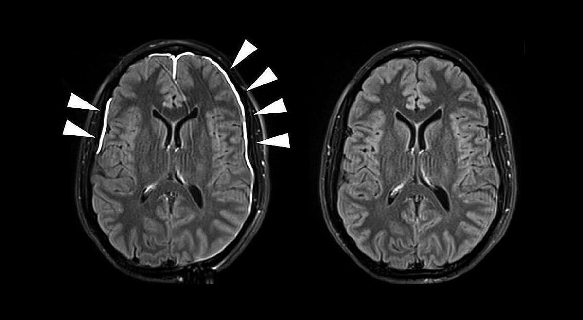
A concussion is defined as a traumatic brain injury (TBI) caused by a bump, blow, or jolt to the head or by a hit to the body that causes the head and brain to move rapidly back and forth. This sudden movement can cause the head and brain to bounce around or twist in the skull, stretching and damaging brain cells and creating chemical changes in the brain. A concussion is generally referred to as a mild traumatic brain injury or mTBI because is usually not life-threatening.
Often referred to as a “silent epidemic”, with an incidence of over 6 million cases/year in the US, mTBIs are complex to diagnose: they rely on self-reported symptoms that may appear sometimes after the trauma. However, due to the risks related to exposing traumatized patients again, sports physicians and associations have increasingly been looking at ways to objectivize the assessment of patients.
Relevance of eye tracking in Concussion
The relationship between eye movements and mTBI has mainly been studied from a clinical perspective. In the literature review below, we shortlisted publications showcasing the relevance of studying eye movements to quickly assess and follow up on traumatized patients in a quantified way. The first article shows promises of eye-tracking for detection in the sports field, the second article for the follow-up during the consultation, and the third article shows the value of eye movements together with the gold standards for follow-up.
1. Fixation of eye movements following concussion
Researchers found that fixational saccade amplitudes were significantly different in the concussion group, but only when fixating on the center of the raster. This task specificity suggests that task optimization may improve differentiation and warrants further study.
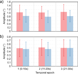
The concussion group is shown in red and control in blue; error bars are ±SD. Figure (a) shows the results for the center fixation tasks while figure (b) shows the results for the peripheral fixation tasks.
Fixational eye movement measured in the acute-to-subacute period of concussion recovery may provide a quick (<3 minutes), objective, sensitive, and accurate ocular dysfunction assessment. Future work should assess the impact of age, mechanism of injury, and post-concussion recovery on FEM alterations following concussion.
Evaluation of concussion requires assessments in multiple domains because the clinical profiles are heterogeneous. While most individuals show recovery within a week of injury, others experience prolonged recovery periods. Visual tracking performance metrics may serve as a biomarker of debilitating symptoms of concussion implicating attention as a root cause of such pathologies
2. Association of visual tracking metrics with post-concussion symptomatology
Results from researchers suggest that visual tracking performance metrics reflect clinical symptoms when assessed within 2 weeks of concussion.
3. Follow-up evaluation of oculomotor performance with fMRI in the subacute phase of concussion
Recent studies show that three of the 7 oculomotor tasks (antisaccade, self-paced saccade, and memory-guided saccade) administered showed significant differences between the recently concussed group compared with normal volunteers.
Even at 30 days postinjury, and despite being clinically asymptomatic, advanced techniques are able to detect subtle lingering alterations in the concussed brain. Therefore, progressive neuroimaging techniques such as fMRI in conjunction with assessment of oculomotor performance may be beneficial in the clinical management of concussion.
You want to know more?
Our experts have gathered a comprehensive list of scientific literature about the relevance of eye tracking in Concussion. Feel free to contact us a request a copy.