


Exploring Antisaccades and Frontal Lobe Function
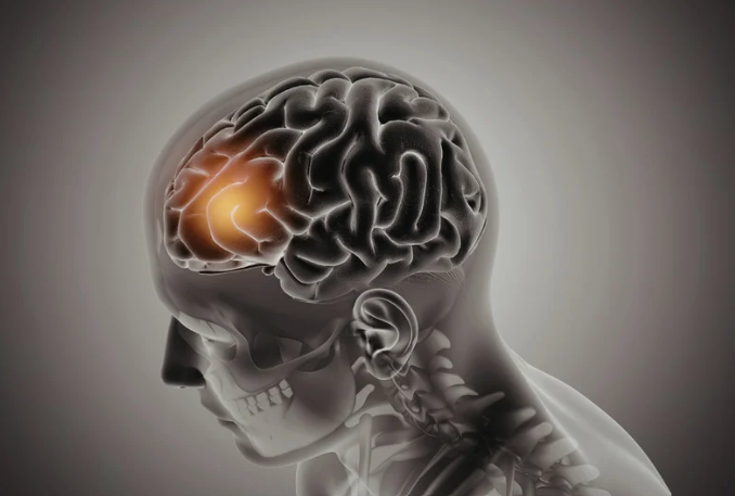
Exploring Antisaccades and Frontal Lobe Function
May 6, 2025 11:00:00 AM
4
min read
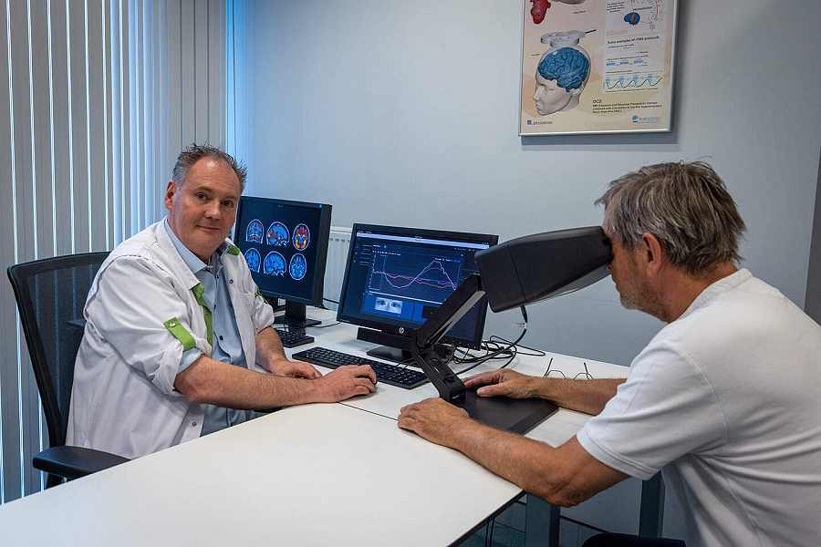
Oculomotor Biomarkers Recorded on 50 Patients with neuroClues®, now CE-Marked!
.png)
Oculomotor Biomarkers Recorded on 50 Patients with neuroClues®, now CE-Marked!
Mar 4, 2025 9:45:00 AM
4
min read
Prof. Dr. Lafosse and RevArte Featured in Belgian Media for Integrating neuroClues® in Neurorehabilitation
Prof. Dr. Lafosse and RevArte Featured in Belgian Media for Integrating neuroClues® in Neurorehabilitation
May 26, 2026 3:08:06 PM
2
min read

